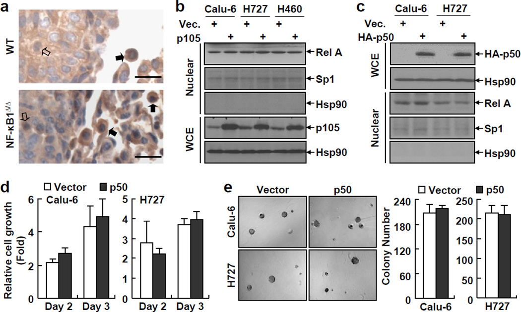
Figure 4.
RelA activation in lung cancer cells is independent of NF-κB1 and expression of exogenous p50 has no effect on the tumorigenicities of NF-κB1low lung cancer cells. (a) IHC staining showing a similar RelA nuclear expression in NF-κB1Δ/Δ tumors and WT tumors from urethane-treated mice. Scale bar: 20 µm. Representatives of tumor cells and immune cells with positive staining of nuclear RelA are indicated by open and filled arrows, respectively. (b) IB assays showing no change in RelA nuclear expression in p105 human lung cancer stable cell lines. (c) IB assays confirming expression of exogenous HA-tagged p50 in human lung cancer stable cell lines and showing no change in RelA nuclear expression in these stable cell lines. (d) Cell growth assays showing no change in the growth of p50 human lung cancer stable cell lines. Data shown are means ± SD (n ≥ 3). (e) Soft agar colony formation assays showing no change in anchorage-independent growth of p50 human lung cancer stable cell lines. Data shown are means ± SD (n ≥ 3).

