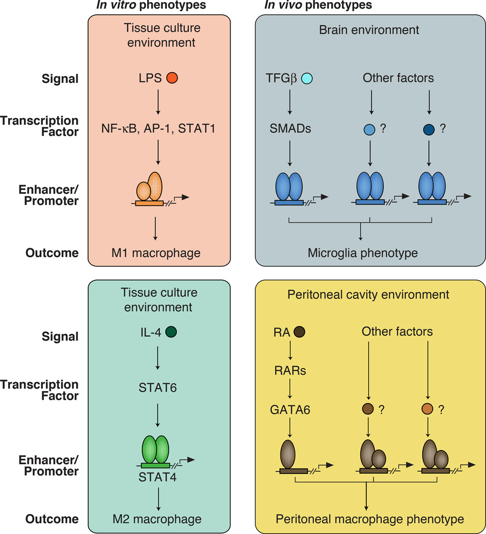
Figure 1. Different signaling pathways in macrophages lead to diverse phenotypic outcomes.
Left panels: Macrophages can be polarized toward M1, M2 phenotypes in in vitro by exposure to LPS or IL-4 respectively.
Right panels: The brain and peritoneal cavity contain high levels of TGFβ or retinoic acid (RA) respectively, that are important determinants of the distinct phenotypes of microglia and resident peritoneal macrophages. However, these are only a subset of what are as yet mostly unknown signals that must be integrated to establish tissue-specific gene expression signatures.

