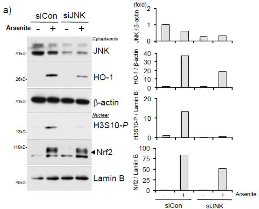
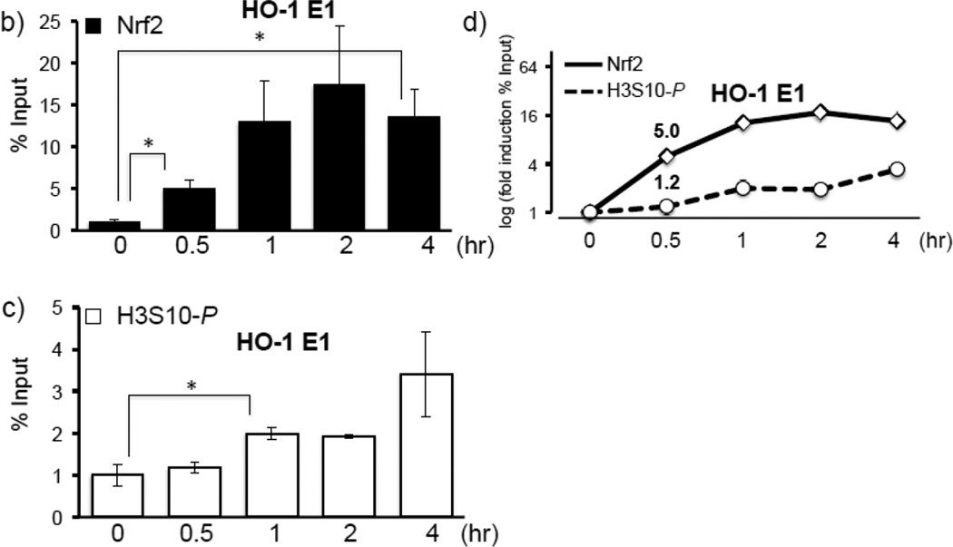
Figure 6. Arsenite induces JNK-dependent H3S10 phosphorylation and recruits Nrf2 to the HO-1 ARE prior to enrichment of phosphorylated H3S10.
a) HaCaT cells were transiently transfected with JNK specific siRNA or control siRNA as described in Materials and Methods. After 48 hr, cells were treated with 10µM arsenite for 8 hr and nuclear and cytoplasmic fractions were subjected to Western blot analysis with anti-HO-1, anti-Nrf2, anti-JNK, and anti-H3S10 phospho-specific (H3S10-P) antibodies; Lamin B and β-Actin blots are shown as loading controls. Quantification of blots is shown to the right. Densitometry analysis was conducted with Image J software. Relative density of arsenite treated signal bands were normalized to the corresponding control signal bands (siCon/arsenite (−)) for fold change, then further normalized to the relative density of loading control signal bands for loading accuracy; JNK and HO-1 were normalized to β-Actin; H3S10-P and Nrf2 were normalized to Lamin B. (b and c) HaCaT cells were treated with 10µM arsenite for 0.5, 1, 2, or 4 hr, then harvested for ChIP assay and incubated with rabbit IgG, anti-Nrf2 (b), or anti-H3S10 phospho-specific (H3S10-P) (c) antibody. Isolated genomic DNA was subjected to quantitative RT-PCR using primer pairs for the HO-1 E1 region. Samples were normalized to input and presented as percent input. Significance was calculated using a t-test and established with p < 0.05. A representative of three independent experiments is shown. (d) Graphical representation of temporal induction of Nrf2 and phosphorylated H3S10 (H3S10-P) enrichment on the HO-1 E1 ARE.


