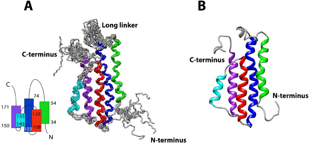
Figure 1. Solution structures of DBPB.
(A) Ensemble of the 10 lowest-energy DBPB structures. Helix 1, consisting of residues 34 to 54, is colored green. Helix 2, consisting of residues 74 to 103, is colored blue. Helix 3, consisting of residues 108 to 128, is colored red. Helix 4, consisting of residues 133 to 143, is colored cyan. Helix 5, consisting of residues 150 to 171, is colored purple. The topology of DBPB is shown at the bottom left. (B) Ribbon depiction of a representative DBPB structure.

