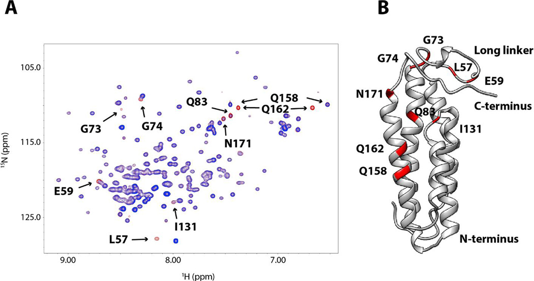
Figure 7. PRE perturbation of DBPB by paramagnetically labeled DS dp12.
(A) 1H- 15 N HSQC overlays of WT B31 DBPB with 8 molar equivalents of TEMPO-labeled DS dp12. HSQC spectrum before the radical is reduced is shown in blue. HSQC spectrum of the protein after reduction of the radical is shown in red. Residues showing prominent PRE perturbations are indicated. They are L57, E59, G73, G74, Q83, I131, Q158, Q162 and N171. (B) Ribbon representation of DBPB in the same orientation as in Figure 4B with TEMPO-perturbed residues colored red.

