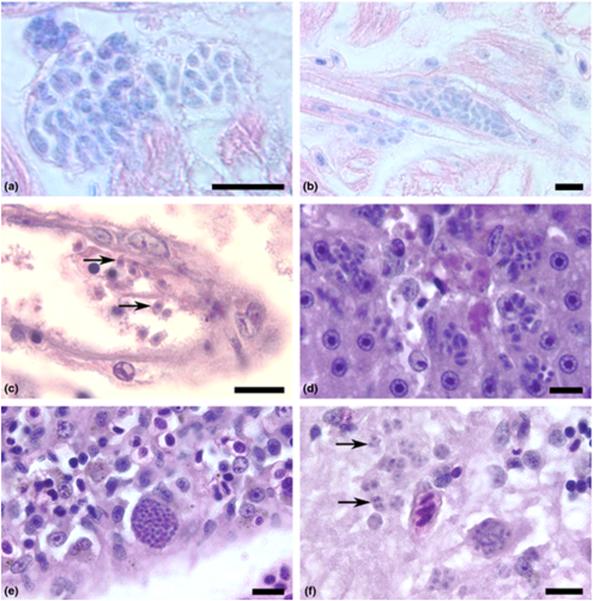
Figure 1.
Tachyzoites of Toxoplasma gondii developing in tissues of zebrafish raised at 37 °C. Haematoxylin and eosin stain. Bar = 10 μm. (a) Two foci of tachyzoites developing within parasitophorous vacuoles in the heart after 6 days post-injection (dpi). Giemsa stain. (b) Tachyzoites developing within a cardiac myocyte. 6 dpi, Giemsa stain. (c) Several extracellular, crescent-shaped tachyzoites (arrows) in the lumen and developing within the endothelium of a small blood vessel of an adult zebrafish after 7 dpi. Haematoxylin and eosin (HE) stain. (d) Several tachyzoites proliferating in the liver parenchyma associated with necrosis. Adult zebrafish at 6 dpi. HE stain. (e) A large pseudocyst filled with developing tachyzoites in the spleen. Adult zebrafish, 6 dpi. HE stain. (f) Numerous tachyzoites (arrows) developing in the grey matter of the midbrain of an adult zebrafish, 6 dpi. HE stain

