
Fig. 2.
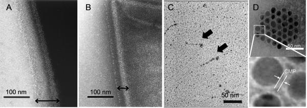
Transmission electron micrographs of B. anthracis spores after negative staining (A and B, reproduced with permission from Ref. [43], © American Society for Microbiology), Scl1 protein after rotary shadowing (C, reproduced with permission from Ref. [44], © American Society for Biochemistry and Molecular Biology), and gold nanoparticles with self-assembled CMP monolayers (D). B. anthracis spores show densely packed collagen-like layers on their surfaces (double headed arrows): Strain 7702 which has 8 copies of (GPT)5GDTGTT sequence displays a thick layer (A), while strain 5725R with only 1 copy of the same sequence displays thin collagen like layer (B). Sc1 protein (streptococcal collagen like protein with charged amino acids) exhibits lollipop morphology similar to complement factors (C, block arrows). Self-assembled CMP layer provides colloidal stability to gold nanoparticles (D).