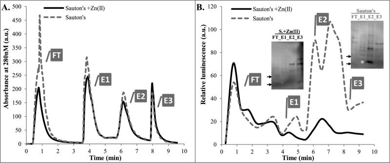
FIGURE 6. Ribosome isolation from M. tuberculosis mc26206 grown in Sauton's medium with or without added Zn2+.
A. FPLC UV traces of two lysates from cultures grown with or without Zn2+, as indicated. FT fraction from growth in Sauton's medium without Zn2+ was pink, indicating presence of mCherry. BugBuster extraction buffer alone strongly absorbs at 280nm in FT (data not shown).
B. ELISA of FPLC fractions using anti-S18-2 antibodies: Each fraction was diluted 4× in 4M urea/TBS, proteins were coupled to wells, and then processed as described in Experimental Procedures.
Insert: Proteins from each peak fraction were precipitated with TCA (20%, overnight at 4°C), washed with cold ethanol, and solubilized in urea/CHAPS buffer. 12μg of each sample was analyzed by Western blot. * band corresponding to MW of the S18-2 protein. Arrows depict 10kDa and 15kDa markers.
The western blot of TRIzol-purified proteins resulted in a smaller number of non-specific bands (see insert in Figure 1D) than sonication in BugBuster buffer (panel B).
FT- flow-through, E1, E2, E3- elution peaks

