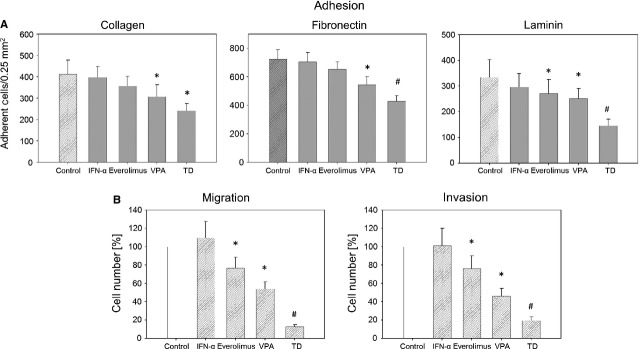
Figure 3.

(A) Adhesion of prostate cancer cells to extracellular matrix proteins. PC-3 cells were treated with 100 U/ml IFNα, 1 nM everolimus or 1 mM VPA, or with all compounds simultaneously (TD). Cells were added to immobilized collagen, laminin or fibronectin at a density of 0.5 × 10⁶ cells/well for 60 min. Plastic dishes were used to evaluate unspecific binding (background control). One representative of six experiments is shown. * indicates significant difference to controls, # indicates significant difference to single drug treatment. (B) PC-3 cell migration (left) and invasion (right). Cells treated with 100 U/ml IFNα, 1 nM everolimus or 1 mM VPA, or with all compounds simultaneously (TD). Controls were set to 100%. * = significant difference to controls. # = significant difference to single drug treatment.
