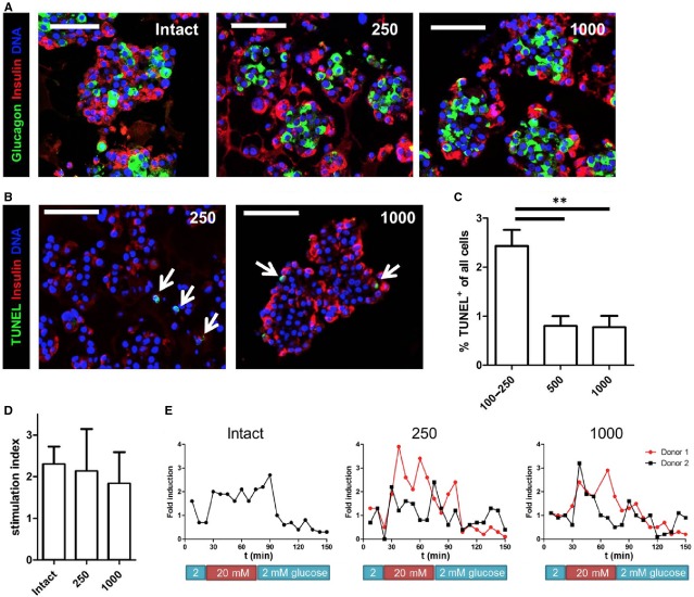
Figure 5.
Human islet cell aggregates are viable and function in vitro. (A) Human islet cell aggregates of all sizes show an architecture with beta cells (red) located at the periphery and alpha cells (green) in the core. In comparison, intact human islets show a heterogeneous distribution of alpha- and beta cells. (B) TUNEL staining showing only few apoptotic cells in both small (250 cells/aggregate) and large (1000 cells/aggregate) human islet cells aggregates (positive cells are indicated with an arrow). (C) Quantification of TUNEL+ cells shows that 2.5% of all cells are apoptotic in small aggregates. Aggregates of 500 or 1000 cells contain less than 1% apoptotic cells (n = 3–4 donors per condition). (D) Static glucose-stimulated insulin secretion (GSIS) test was performed on human islet cell aggregates of 250 and 1000 cells and compared with intact control islets. Islet cell aggregates of both sizes show a similar secretory response as intact control islets; error bars represent ±SD. (E) Dynamic GSIS shows more sustained insulin secretion during high glucose in intact islets compared to human islet cell aggregates, although the induction is similar; scale bar = 100 μm.

