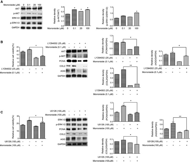
Figure 2.
Morroniside activates AKT and ERK in human OA chondrocytes. (A) Cells were treated with different dose of morroniside (0.1, 20 and 100 μM) for 24 hrs. The levels of AKT, ERK, p-AKT and p-ERK were detected by western blotting analysis. (B) Cells were pre-treated with LY294002 (25 μM) for 2 hrs prior to treatment with morroniside (0.1 μM) for 24 hrs. The cell viability was measured by the MTT assay, and the levels of AKT, p-AKT, PCNA, COL2 and AGG were detected by western blotting analysis, respectively. (C) Cells were pre-treated with U0126 (100 μM) for 2 hrs prior to treatment with morroniside (100 μM) for 24 hrs. The cell viability was measured by the MTT assay, and the levels of ERK, p-ERK, PCNA, COL2 and AGG were detected by Western blotting analysis, respectively. The values represent the mean ± SEM of three to five independent experiments, each yielding similar results (*P < 0.05, **P < 0.01).

