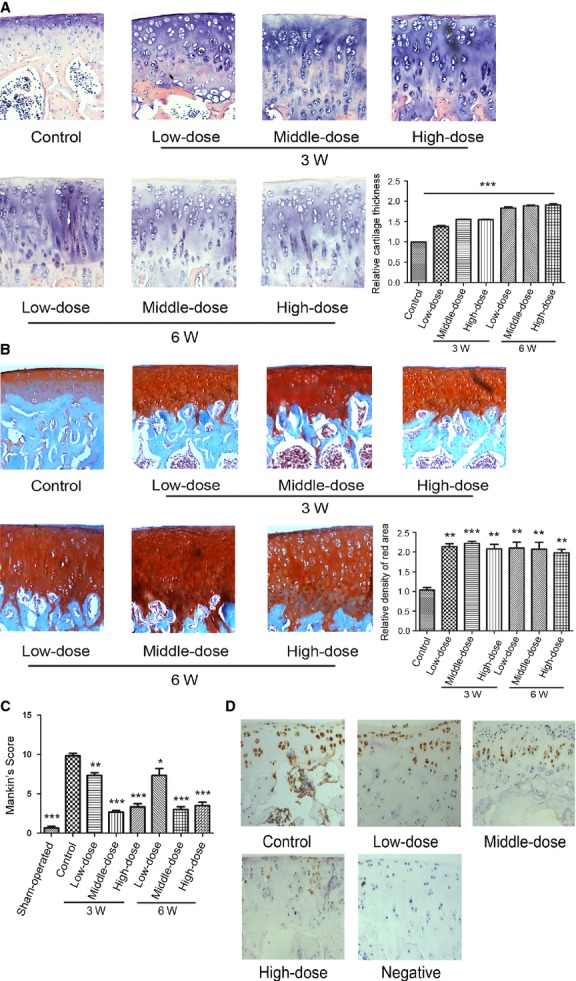
Figure 4.

Morroniside ameliorates the cartilage damage in a rat experimental model of OA Specimens were longitudinally cut into 3–5 μm sections. (A) Sections in different treated groups were examined using haematoxylin and eosin staining (original magnification ×100). (B) Sections in different treated groups were examined using Safranin O-fast green staining (original magnification ×100). The index of matrix production was evaluated by relative density of red area. The values represent the mean ± SEM of three to five independent experiments, each yielding similar results (**P < 0.01, ***P < 0.001). (C) Histopathological scores were performed by Mankin’s score. (D) Sections in morroniside-treated 6-week group were performed with TUNEL assay (original magnification ×100).
