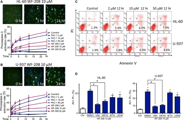
Figure 4.
WF-208 induced HL-60 and U-937 apoptosis through the activation of procaspase-3 to caspase-3. (A) WF-208 and PAC-1 induced the cleavage of procaspase- 3 to caspase-3 in HL-60 cells (upon) and U-937 (below) at different time and concentration. *P < 0.05 versus control group. (B) WF-208 and PAC-1 induced the cleavage of procaspase- 3 to caspase-3 in U-937 cells. The representative image of WF-208 (upon) and activation rate of WF-208 and PAC-1 (below) induced procaspase-3 cleavage in U-937 cells at different time and concentration. *P < 0.05 versus control group. (C) Phosphatidylserine exposure (measured by Annexin V/PI co-staining, describe as the percentage of Annexin V-positive PI-negative cell percentage) in HL-60 and U-937 after 24 h treatment with 2, 10, 50 μM WF-208. (D) The inhibition of 50 μM vancaspase inhibitor (Z-VAD-FMK), caspase-3 inhibitor (Z-DEVD-FMK), caspase-8 inhibitor (Z-LETD-FMK), caspase-9 inhibitor (Z-LEHD-FMK) on early apopsotis induced by 10 μM WF-208 in HL-60 and U-937 cell lines treatment 24 hrs. *P < 0.05 versus control, #P < 0.05 versusWF-208 alone.

