Figure 1.
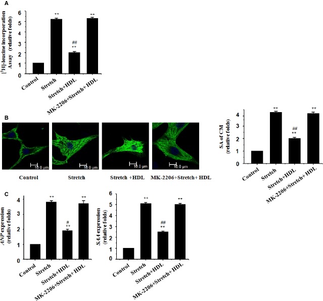
HDL inhibits mechanical stress-induced hypertrophic responses in cultured cardiomyocytes. Cultured rat neonatal cardiomyocytes were treated with vehicle (control) or mechanical stretch in the absence or presence of HDL (100 μg/ml), MK-2206 (100 nM) or both; (A) [3H]-Leucine incorporation in cardiomyocytes; mean ± SEM from 3 independent assays. (B) Cardiomyocyte morphology and size in cardiomyocytes subjected to immunofluorescence staining for α-MHC (green) and DAPI. Representative photographs from 3 independent experiments are shown (scale bar: 10 μm). The surface area (SA) of cardiomyocytes was evaluated by measuring 100 cardiomyocytes in each dish. (C) Expression of the ANP and SAA genes evaluated by real-time RT-PCR; β-actin was the internal control.

