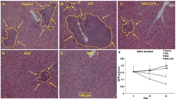
Figure 3.

Lymphocytic infiltrates in salivary glands of NOD mice. A–E: H&E staining showing lymphocytic infiltration size (shown by yellow line and arrows) in NOD that were: untreated (A), CFA-treated (B), MSC + CFA (C), MSC (D). In 2 of the 5 NOD mice transplanted with MSCs only, no lymphocytic infiltrates were noted (E). Scale bar: 140 um for all images. (F): Salivary flow rates (SFRs) of NOD mice. SFRs in MSC + CFA (black circle; n = 10) and MSC (black square; n = 5) groups did not decrease during the follow-up period (22 wk of age) and were significantly higher than SFRs of CFA-treated or control NOD groups (n = 5 per group; P < 0.05). SFRs in CFA (triangular) or control (untreated; open circle) groups continued to decrease during the follow-up period (*P < 0.05). Reproduced with permission from: [35].
