
Fig 2. Overexpression of miR-29a decreased GFAP, a marker of HSC, and increased expression in the acute response to injury and immunoreactivity in cholestasis.
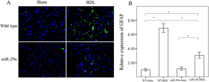
There was significantly higher expression of GFAP (green) in tissues from the BDL group than in tissues from the sham-operated group in WT mice. Moreover, miR-29a overexpression significantly downregulated GFAP protein expression in miR-29aTg mice with cholestasis compared with the WT littermates. Data are expressed as the mean ± SE of six samples per group. *indicates a p < 0.05 between the groups.