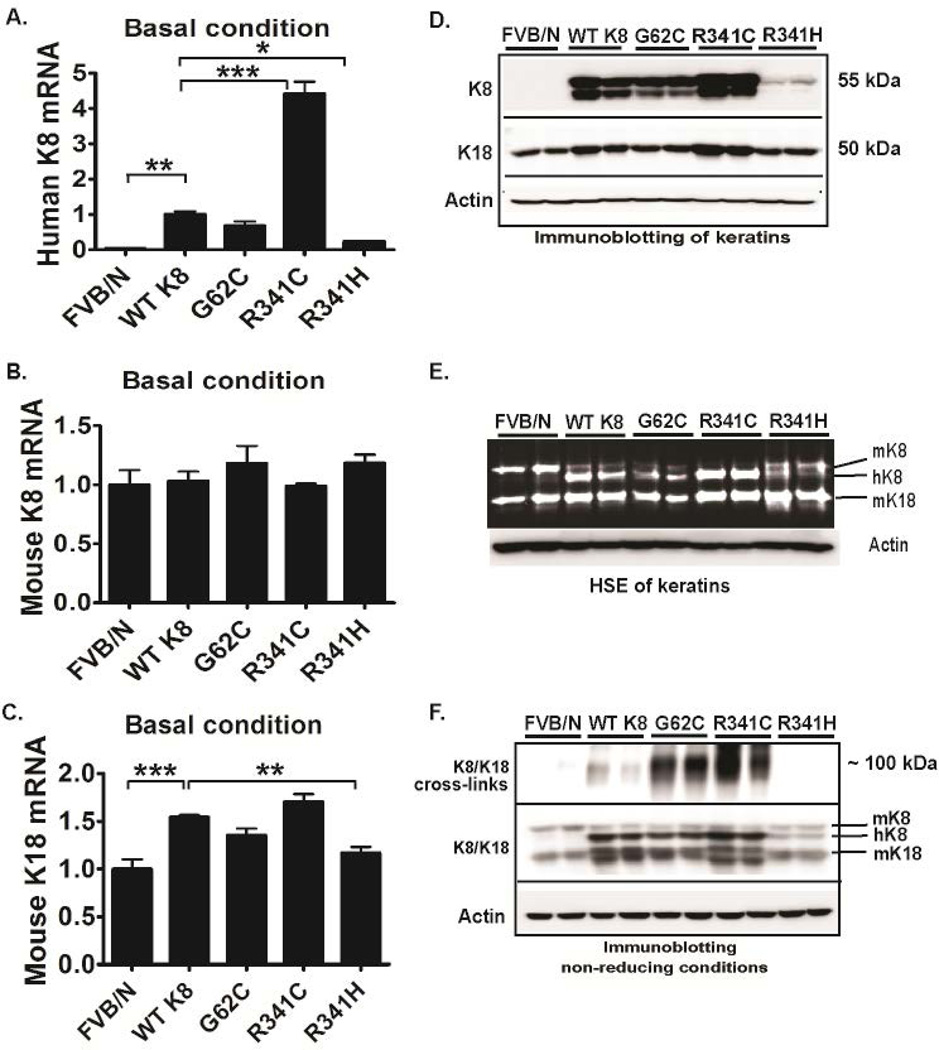
Figure 1. Keratin 8 G62C/R341C variants promote keratin crosslinking under basal conditions.
Human (h) K8 (A) and mouse (m) K18/K8 (B,C) mRNA and protein levels (D) were quantified by RT-PCR and immunoblotting, respectively. (E) To directly visualize proteins, a high salt extraction with SYPRO ruby staining was performed from nontransgenic livers (FVB/N) as well as tissues overexpressing wild-type K8 (WT K8) or the highlighted K8 variants. (F) To test the formation of disulfide bridges, livers were homogenized under non-reducing conditions. After longer exposure (upper panel), significant amount of cross-linked hK8 was detected in G62C and R341C livers and the crosslinks went away under reducing conditions (not shown). L7 (mouse ribosomal protein) and actin were used as an internal/loading control for qRT-PCR and immunoblotting, respectively. At least four mice were analysed per genotype and the results are expressed as mean ± SEM. *p<0.05, **p<0.01, ***p <0.001

