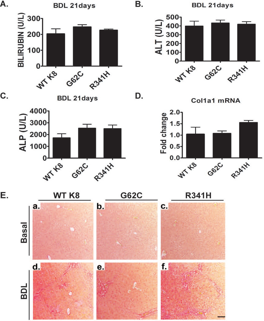
Figure 7. Presence of K8 variants does not affect the development of bile duct ligation (BDL)-induced liver fibrosis.
(A–C) Three weeks after BDL, mice displayed significantly elevated bilirubin, ALT and alkaline phosphatase (ALP) levels, however no significant differences were noted among the genotypes. Furthermore, In all untreated animals, bilirubin, ALT and ALP levels were within normal range. (E) The extent of liver fibrosis in untreated animals (basal) as well as mice subjected to BDL for three weeks was evaluated by picro-sirius red staining. Note that animals overexpressing WT K8 or the depicted K8 variants display a similar amount of liver scaring and similar hepatic collagen mRNA levels as determined by qRT-PCR (D). L7 (mouse ribosomal protein) gene was used as an internal control. At least four mice were analysed per genotype and the results are expressed mean ± SEM. Scale bar=100µm.

