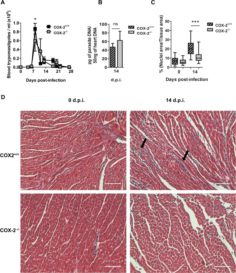
Fig 3. Parasite burden and heart inflammation in T. cruzi infected COX-2+/+ and COX-2-/- mice.
(A) The presence of parasites in the blood of COX-2+/+ or COX-2-/- mice at different d.p.i. was quantified by direct counting under optical microscopy. (B) DNA from heart tissue was isolated and qPCR using T. cruzi DNA standard was performed to determine parasite burden in COX-2+/+ or COX-2-/- infected mice at 14 d.p.i. Means ± SEM from three independent experiments are shown (n = 4). (C) Heart tissue sections of COX-2+/+ and COX-2-/- mice either non-infected (0 d.p.i.) or 14 d.p.i., were stained with Masson’s Trichrome and inflammatory cell infiltration was quantified as described in Methods. (D) Representative pictures of heart tissue sections described in C. Arrows indicate inflammatory infiltration. Scale bar is 100 μm. (ns = non-significant; *p<0.05; **p< 0.01; ***p<0.001).

