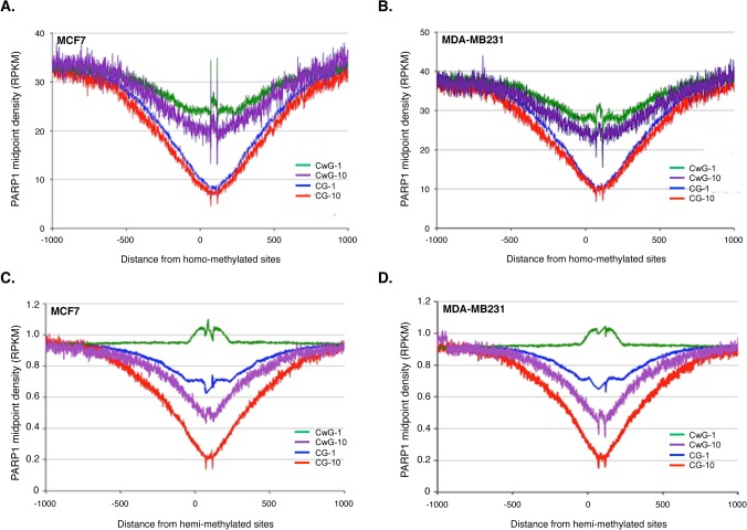
Fig 4. Global analysis of PARP-1 association with methylated DNA.
Genome-wide methylated CpG and non-CpG sites were aligned with PARP-1-associated nucleosomal DNA sequences obtained from PARP1-nuc-ChIP-seq and showed mutually exclusive pattern between both signals. (A) PARP1-binding at methylated sites of MCF7 cells (B) MDA-MB231 cells. CG-1 and CG-10 (blue and red) lines are PARP-1 association with moderately and highly methylated sites respectively. CpG sites; CwG-1 and CwG-10 (green and violet) represent PARP1 presence at moderately and highly methylated non-CpG sites (see methods). (C) PARP1 presence at hemi-methylated DNA in MCF7 cells and (D) in MDA-MB231 cells. CG-1 and CG-10 (blue and red lines) indicate moderately and highly hemi-methylated sites while CwG-1 and CwG10 represent moderately and highly hemi-methylated non-CpG sites respectively.

