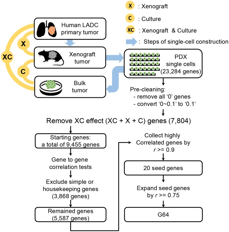
Fig 1. Schematic illustrating the preparation of coordinately co-expressed genes across the 34 single LADC cells.
The upper part of the schematic illustrates the sequential procedures of LADC tissue isolation, mouse engraftment, patient-derived xenograft (PDX) cell culture, and single PDX cell preparation for RNA sequencing [28]. The lower part of the schematic explains how the coordinately expressed genes were selected from the 34 single-cell transcriptomes. Please refer to the Materials and Methods section for a detailed description of the procedures.

