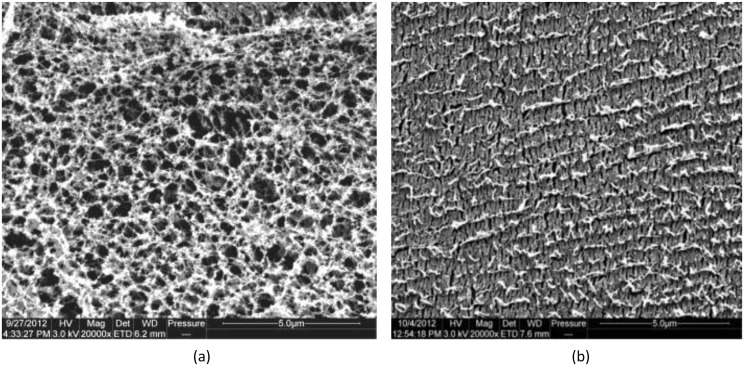
Fig 3. Cryo-Scanning electron micrograph images of unstimulated SMSL and parotid saliva.
(a) section of unstimulated SMSL saliva showing the appearance of a filamentous mucin network; (b) section of parotid saliva showing a continuous matrix with the absence of a filamentous mucin network (Scale bar = 5μm).

