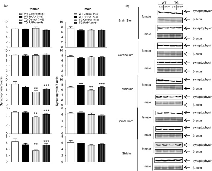
Fig. 3.
Effect of RAPA on synaptophysin protein level in midbrain, striatum, brain stem, cerebellum, and spinal cord. (a) quantification of Western blot; (b) representative immunoblots. Age-matched female WT and TG mice were fed mouse diet incorporated with microencapsulated RAPA and the microencapsulation material Eudragit S100 (vehicle control) from 13 weeks of age for 24 weeks. Each well of a 4–12% Criterion gel was loaded with 40 µg brain tissue lysate. Immunoreactive bands were quantified by Odyssey software. Data represent the mean±SEM. Two-way ANOVA with post hoc Bonferonni t-tests were used to analyze the effects of genotype and RAPA on levels of synaptophysin in either sex. *p<0.05, WT control versus WT RAPA; **p<0.05, WT control versus TG control; ***p<0.05, TG control versus TG RAPA.

