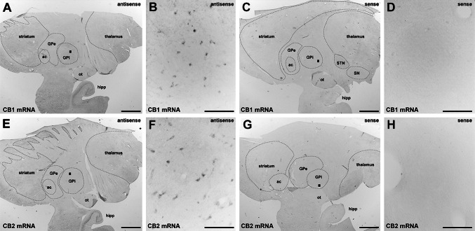
Fig. 2.
Detection of CB1R and CB2R mRNA using in situ hybridization. Using colorimetric in situ hybridization in a naïve primate, CB1R and CB2R mRNA (panels a, b and e, f, respectively) were detected in the GPi nucleus. The sense probes did not provide specific labeling of CB1R or CB2R mRNA (panels c, d and g, h, respectively). Even at low magnification, a lack of stain when using sense probes for CB1R and CB2R mRNA (panels c and g) was observed in the hippocampal formation, which was stained specifically when using antisense probes for both transcripts (a and e). Scale bar is 3,000 μm for panels a, c, e and g and 150 μm for insets b, d, f and h. ac anterior commissure, GPe external division of the globus pallidus, GPi internal division of the globus pallidus, hipp hippocampal formation, ot optic tract, SN substantia nigra, STN subthalamic nucleus

