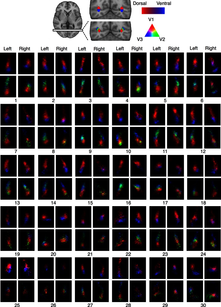
Fig. 7.
Visitation maps of optic radiation tractography in all thirty participants tested. All maps sampled at a single coronal slice immediately posterior to the lateral ventricle. Visual field-based segmentation shows the superior–inferior division of streamlines, in red and blue. Hierarchy-based segmentation discriminates streamlines into dorso–lateral nesting compartments, in red, green and blue

