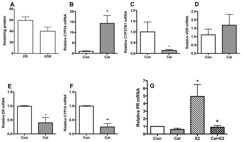
Fig. 1. Presence and functional activity of ER and VDR in tissue slice cultures of MMTV-Wnt1 tumors.
MMTV-Wnt1 tumor orthografts were used to generate tissue slices. The slices were cultured and the expression of ER and VDR and their functional responses were determined as described in Methods Section. A, Basal ER and VDR levels. [3H]-labeled ligand binding assays revealed the expression of ER and VDR proteins in the tumor slices. B–F, VDR functional responses. Tissue slice cultures were treated with 0.1% ethanol vehicle or 100 nM calcitriol (Cal) for 5 h and the mRNA levels of the Cyp24, Cyp27B1, Vdr, Erα and Cyp19 were determined by qRT-PCR (n = 4; * p<0.05 and ** p<0.01 as compared to the Std group). G, ER functional response. Tissue slice cultures in phenol red-free culture media were exposed to 0.2% ethanol vehicle (control, Con), 100 nM calcitriol (Cal), 10 nM E2 (E2) or a combination of both (Cal+E2) for 5 h and PR mRNA levels were determined by qRT-PCR (n = 4; * p<0.05 as compared to Con and + p < 0.05 as compared to E2). At least three different orthografts were used to generate the tissue slices and each experiment was conducted in duplicate. Values represent mean ± SEM.

