Figure 1.
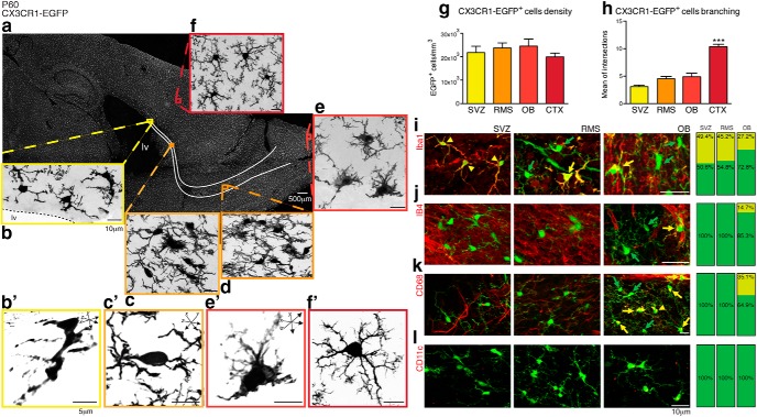
CX3CR1-EGFP+ cells displaying a distinct morphology comprise a component of the adult SVZ/RMS/OB. Confocal microscopy analysis of brain parasagittal section obtained from adult CX3CR1-EGFP mice (P60) reveals that microglia (EGFP, gray) are distributed throughout the brain (a) and also in the SVZ (b), along the RMS (c, d) and OB (e). Microglia within the SVZ niche have enlarged cell bodies and display an immature morphology (b′, c′, e′). Despite their distinct morphology compared with their counterparts in the cortex (CTX), in which typical ramified microglia are observed (f, f′), the cell density is similar in all analyzed regions (SVZ: 22 × 103 ± 2.8 × 10³; RMS: 24 × 103 ± 2.2 × 10³; OB: 24.5 × 10³ ± 3 × 10³; CTX: 20 × 103 ± 1.7 × 10³; mean ± SEM; p > 0.05, Tukey's multiple-comparisons test; g). Along the SVZ niche, Scholl analysis of cell branching shows that microglia are markedly less ramified than their counterparts distributed in the cortical parenchyma (SVZ: 3.01 ± 0.3; RMS: 4.5 ± 0.4; OB: 4.9 ± 0.6; CTX: 10.3 ± 0.5; mean ± SEM of intersections to concentric circles; p = 0.0001, Kruskal–Wallis test; h). Analysis of brain sections obtained from CX3CR1-EGFP transgenic mice (P60) and stained for Iba1 (i, red), Isolectin B4 (j, red), and CD68 (k, red) show that the majority of CX3CR1-EGFP+ cells (green, indicated by green arrows) are not colabeled in the SVZ, RMS, or OB (few double-labeled cells are indicated by yellow arrows). Despite the lack of immunoreactivity to these markers, CX3CR1-EGFP+ cells present in the SVZ/RMS/OB do not appear to be dendritic cells, as they do not express CD11c (l). Scale bars: a–f, i–l, 10 μm; b′, c′, e′, f′, 5 μm.

