Figure 2.
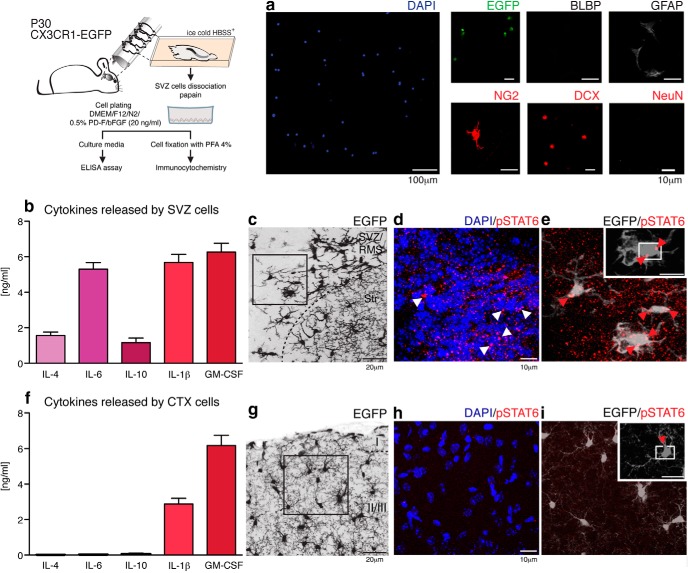
The adult SVZ is enriched in cytokines that promote neurogenesis. The schematic depicts the culture of SVZ cells. These were plated at density of 1–3 × 10⁵ cells/ml and their phenotype was assessed by immunocytochemistry. Dissociated cells correspond to microglia (EGFP, green), astrocyte/astrocyte-like stem cells (GFAP, gray), cells of the oligondendroglial lineage (NG2, red), and neuroblasts (DCX, red). BLBP+ astroglia and mature neurons (NeuN+) were not observed (a). Shortly before fixation, culture media were collected and the cytokine-release profiles of SVZ cells were assessed by ELISArray assay. IL-1β (5.7 ± 0.5 ng/ml), IL-4 (1.6 ± 0.2 ng/ml), IL-6 (5.3 ± 0.4 ng/ml), IL-10 (1.2 ± 0.2 ng/ml), and GM-CSF (6.3 ± 0.5 ng/ml) were released by mature SVZ cells (b). In contrast, only IL-1β (2.9 ± 0.3 ng/ml) and GM-CSF (6.2 ± 0.6 ng/ml) were released at detectable levels by cortical cells (f). IL-1α, IL-2, IL-5, IL-12a, IL-13, IL-17a, and G-CSF were either not released or released at undetectable levels. The presence of IL-4 and IL-10 led us to investigate whether SVZ/RMS CX3CR1-EGFP+ cells corresponded to alternatively activated-microglia, by immunostaining for pSTAT6, the active form of STAT6 implicated in the determination of this phenotype. c, CX3CR1-EGFP+ cells surrounding the lateral ventricles (EGFP, gray) exhibit a nuclear distribution (counterstaining by DAPI, blue) of pSTAT6 (red) (d, e, arrowheads). In contrast, cortical (CTX), ramified microglia (g) show only rare staining (h, i, red arrowhead). Scale bars: a, 100 and 10 μm; c, g, 20 μm; d, e, h, i, 5 μm.

