Figure 3.

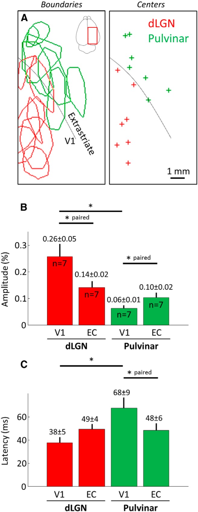
Properties of the cortical responses evoked by thalamic stimulation. A, Left, Delimitations of the cortical surfaces activated in response to dLGN (red) and pulvinar nuclei (green) stimulation from all animals tested. Data originating from both hemispheres were pooled. Drawings delimit stimulated cortical areas based on the image immediately following response onset. Right, Crosses indicate centers of each ROI presented on the left. B, C, Amplitude and latencies measured in area V1 and extrastriate cortical areas after pulvinar and dLGN stimulations (±SEM across values and number of observation). *p < 0.05, EC, Extrastriate cortex.
