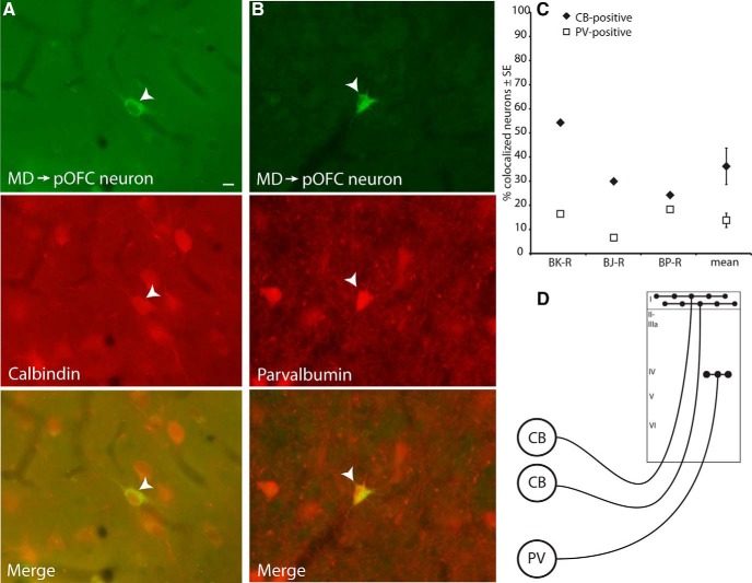
Figure 3.
Neurons in MD projecting to pOFC colocalized with CB or PV. A, Thalamic MD neuron projecting to pOFC (top, white arrowhead) is also positive for CB (middle), as seen in the merged images (bottom). B, Thalamic MD neuron projecting to pOFC (top, white arrowhead) is also positive for PV (middle), as seen in the merged images (bottom). Scale bar in A is 10 μm and applies to A and B. C, Thalamic neurons projecting to pOFC more frequently colocalized with CB than PV, in all cases analyzed. Vertical lines indicate SEM. D, Schematic depicting the termination pattern of calbindin thalamocortical neurons, which target robustly the upper cortical layers, and parvalbumin thalamocortical neurons, which terminate focally in the middle cortical layers.

