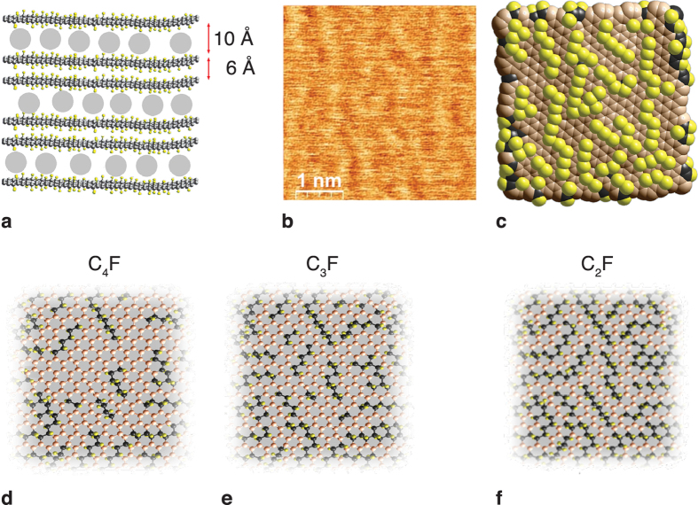
Figure 1. Structure of the CnF compounds.
(a) Side view. The bilayers of fluorinated graphenes are separated by intercalating guest molecules (grey circles). (b) Atomic force microscopy (AFM) topographical image of a basal plane of a C3F sample. (c) Atomistic reconstruction of the AFM image. (d–f) Basal-plane structures of samples with continual C-sp2 nanosegments in C4F (d) and in C3F (e) and isolated C-sp2 nanochains (C2F) (f) plotted according to a set of spectroscopic data17,18.

