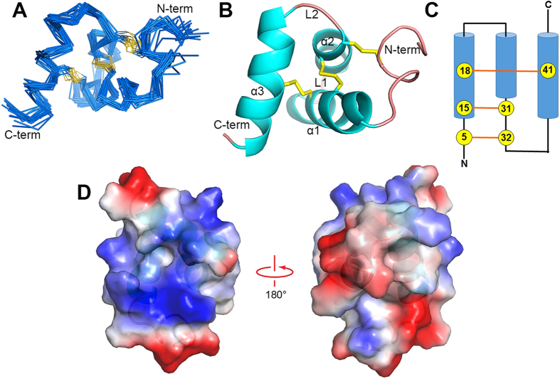
Figure 4. Solution NMR structure of SSD609.
(A) Superposition of the final 20 backbone structures, with the N- and C-termini labeled. (B) Cartoon representation of the SSD609 structure with three disulfide bonds (yellow sticks). (C) Topology diagram of SSD609. Cysteine residues are shown with numbers in circles, and the disulfide bonds are indicated with orange lines. (D) Electrostatic potential surfaces of SSD609.

