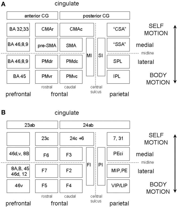
Figure 7.
(A) A highly schematic representation of somatotopically organized sensory and motor cortical areas organized on a medial vs. lateral and frontal vs. parietal oriented map of one hemisphere. The map is bounded medially by the cingulate gyrus and frontally by the prefrontal cortex. The frontal areas are further partitioned according to their rostral or caudal location in the map. The PMC is divided into four regions, labeled PMdc, PMdr, PMvc, and PMvr. The SMA/CMA region is similarly partitioned into CMAc, CMAr, pre-SMA, and SMA. The caudal motor areas each have a corresponding parietal receiving area, which we have labeled the “cingulate sensory area” (CSA), the “supplementary sensory area” (SSA), the SPL, and IPL. (B) Shows the same as (A) but with the equivalent non-human primate labels attached.

