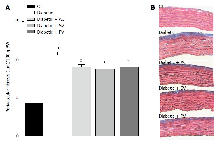
Figure 5.

Representative histological sections of aortic segments from untreated and statin-treated diabetic rats, and untreated control. A: Quantified thickness of perivascular fibrosis in comparable aortic segments from treated diabetic rats and untreated diabetic rats. Perivascular fibrosis was higher in untreated diabetic rats than in CT. All statins decreased perivascular fibrosis in diabetic rats. The values shown are the means ± SEM of 5 animals per group, with the mean value for each animal based on five measurements of its aortic segment. aP < 0.05 for untreated diabetic rats vs untreated CT; cP < 0.05 for untreated diabetic rats vs treated diabetic rats; B: Representative histological sections (× 40, Azan-Mallory stain) of aortic segments from untreated diabetic rats and treated diabetic rats, demonstrating the typical reduction in perivascular fibrosis after treatment with each individual statin. AV: Atorvastatin; SV: Simvastatin; PV: Pravastatin; CT: Control.
