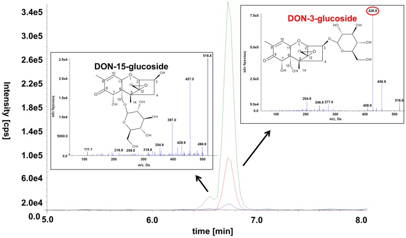
Figure 7.
Chemical structure, SRM chromatograms and MS/MS spectra of the two DON-glucoside isomers in a diluted wheat sample. The following transitions are displayed: m/z 517.1→457.1 (green), m/z 517.1→427.1 (red), m/z 517.1→59.0 (blue). The transition m/z 517.1→427.1 is specific to D3G and can thus be used to distinguish between the two isomers. The MS/MS spectra of the precursor ion at m/z 517.0 [M + Ac]− were recorded at a collision energy of −20 eV.

