Figure 9.

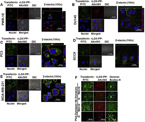
Co‐localization of rLOX‐PP‐Atto565 and transferrin‐FITC in various cell lines and inhibition of rLOX‐PP‐Atto565 (A–E), and 10 kDa dextran‐Bodipy‐fl and transferrin‐FITC uptake in presence or absence of dynasore in PC3 cells at 3 h (F). Cells were incubated with FITC‐transferrin (green) and rLOX‐PP‐Atto565 (red) for 15 min on ice, and then 30 min at 37 °C before imaging. Nuclei were stained with Hoechst 33342 (blue). Confocal microscope images were formatted in split Z‐stacks on the left and merged on the right for each cell lines. PWR‐1E (A), DU145 (B), PC3 (C), SCC9 (D) and MDA‐MC‐231(E) cell lines were listed. Dichromatic (DIC) images are also shown. Transferrin (green) and rLOX‐PP‐Atto565 (red) co‐localization was observed only in PC3 cells and confirms that rLOX‐PP‐Atto565 enters the PC3 cells through a clathrin‐mediated pathway. In (F), uptake of transferrin‐FITC was assessed after 30 min, while that of rLOX‐PP‐Atto565 and 10 kDa dextran‐Bodipy‐fl were assessed after three hours, in the presence or absence of dynasore in PC3 cells with or without quenching of extracellular fluorescence using a Zeiss Axiovert 100M inverted fluorescence microscope.
