
Figure 2.
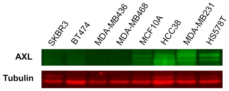
Western blot analysis of Axl protein expression in breast cancer cell lines. Four of the basal-‘B’ triple negative lines we examined (MCF10A, HCC38, MDA-MB231 and HS578T) demonstrated moderate-to-high levels of Axl expression while the MDA-MB436 and MDA-MB468 lines showed no detectable Axl expression. As expected, no Axl protein was detected in the luminal breast cancer cell lines SKBR3 and BT474.