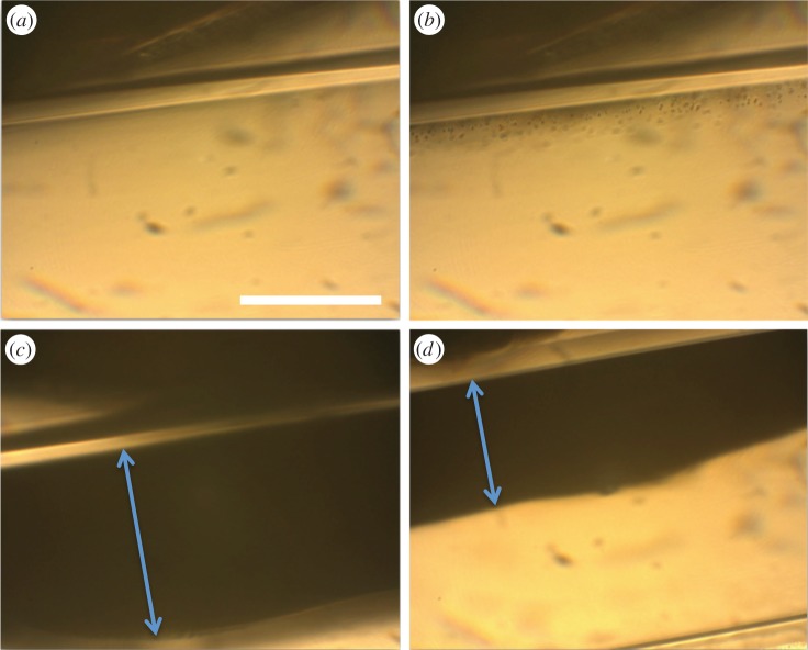
Figure 7.
Optical micrographs showing retention of magnetic microbubbles in a 200 µm inner diameter cellulose tubing adjacent to a permanent magnet providing a magnetic field of 0.37 T and gradient 78.5 T m−1 at the tubing wall. (a) Before injection of microbubbles, (b) immediately following injection of microbubbles at a shear rate of approximately 2100 s−1, (c) 30 s after injection at a shear rate of approximately 2100 s−1 and (d) 30 s after injection at a shear rate of approximately 11 000 s−1 (scale bar indicates 100 µm). The double headed arrows indicate the width of the microbubble bolus formed.

