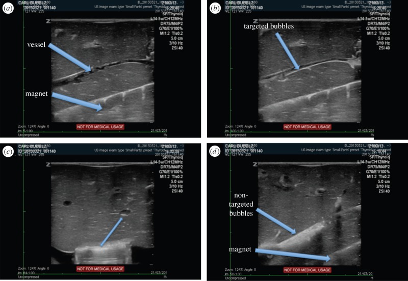
Figure 8.
Ultrasound images showing a blood vessel in an ex vivo perfused liver model with a magnetic Halbach array positioned underneath it. (a) Before injection of magnetic microbubbles, (b) showing magnetic microbubbles retained at the vessel wall, (c) transverse view showing misalignment of the magnetic array and (d) non-magnetic microbubbles flowing through the vessel (second liver).

