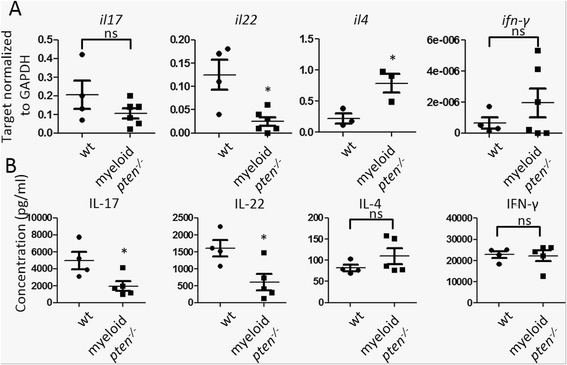
Fig. 5.

PTEN in APCs allows for priming of Th17 cells in vivo. a, b Cells of draining LN of wt (n = 4) and myeloid pten -/- (n = 6) mice were harvested 2 weeks after induction of CIA. a mRNA levels of the indicated cytokines were determined by qPCR. Horizontal bars are means of each group ± s.e. Each data point represents an individual animal *P ≤ 0.05. b LN cells of wt and myeloid pten -/- mice were stimulated in vitro with anti-CD3 for 3 days and levels of the indicated cytokines in the supernatant were measured by ELISA. Horizontal bars are means of each group ± s.d. Each data point represents an individual animal *P ≤ 0.05. APCs antigen-presenting cells, CIA collagen-induced arthritis, ELISA enzyme-linked immunosorbent assay, LN lymph node, PTEN phosphatase and tensin homolog, qPCR quantitative real-time polymerase chain reaction, Th T helper, wt wild-type
