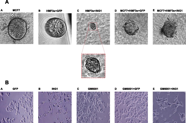
Fig. 5.

Three dimensional co-culture of MCF7 and HMF3s cells. Panel a ING1a induces disorganization of MCF7 breast cancer cell-derived organoids. MCF7 or HMF3s cells were grown individually or co-cultured in Matrigel in three-dimensional cultures in ultralow attachment 96-well plates. Representative images of day 14 (a) MCF7 cells alone (b) HMF3s cells expressing GFP (c) HMF3s cells expressing ING1a; inset magnified spheroid (d) MCF7 cells co-cultured with GFP expressing HMF3s cells (e) MCF7 cells co-cultured with HMF3s cells expressing ING1a captured by digital camera phase contrast microscopy. Panel b MMP blockade partially reverses ING1a induced morphology changes in MCF7 cells. MCF7 cells were supplemented with conditioned media from HMF3s cells infected with Ad-ING1a or Ad-GFP, alone or in combination with GM6001. Representative images after 24 h were captured using a phase contrast microscope
