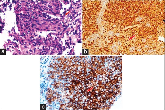
Figure 4.

(a) Large mononuclear pale staining cells with ill-defined cellular margins, Langerhans cells interspersed with inflammatory cells are seen on microscopic examination. (b) Immunohistochemistry showing presence of Langerhans cell S100 protein. (c) Immunohistochemistry showing presence of Langerhans cell CD1a antigen
