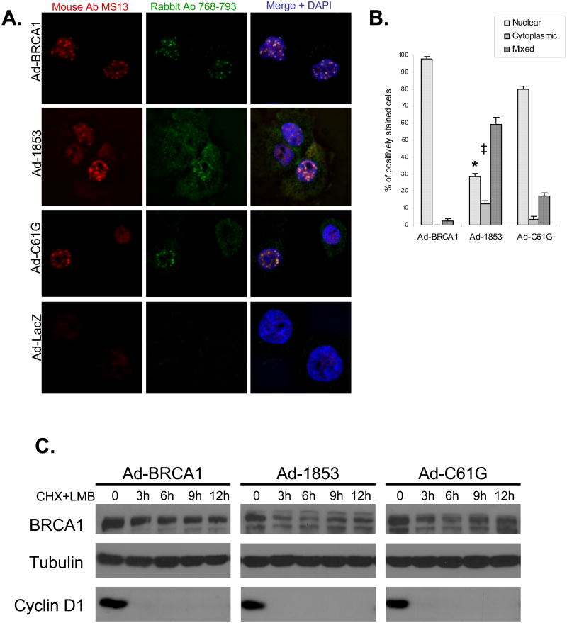
Figure 3. Inhibition of Nuclear Export Promotes the Nuclear Accumulation and Improves the Protein Stability of BRCT-truncated BRCA1.
A) HCC-1937 cells were transduced at MOI=100 with adenoviral vectors expressing the indicated forms of BRCA1 or β-gal (Ad-LacZ) as a control. Cells were cultured for 48 hours to allow for protein expression prior to treatment with 10 nM Leptomycin B (LMB) for 10 hours prior to fixation and immunostaining with two anti-BRCA1 antibodies. Overlap of red and green channels in the merge produces a yellow signal which indicates specific staining of BRCA1. Nuclei are stained with DAPI. Focal nuclear staining of truncated 1853stop BRCA1 protein is increased following LMB treatment in comparison to untreated cells (Figure 2). The experiment was performed independently three times concurrently with three of the five experiments represented in Figure 2. B) Quantification of subcellular localization of wild-type and mutant BRCA1 proteins following LMB treatment. 200 positively stained nuclei were counted for each experiment, N=3, error bars represent SEM. In comparison to untreated cells analyzed at the same time, LMB treatment significantly increased (*, p<0.001) the amount of nuclear and decreased the amount of cytoplasmic (‡, p=0.02) 1853stop BRCA1 protein. Concurrent untreated samples are represented within the composite of Figure 2b. Comparison performed by student's t-test. C) HCC-1937 cells were transduced with adenoviral vectors and cultured for 48 hours. Cells were then pretreated with LMB at 10 nM for 1 hour, and next treated with a combination of 10 nM LMB and 50 μM cycloheximide (CHX) for the indicated time course. Cell lysates were immunoblotted for BRCA1, and densitometry analysis was performed. The t1/2 of 1853stop BRCA1 protein increased to 6.7 hours following LMB treatment in comparison to untreated cells (4.7 hours, Figure 1B). The t1/2 of wild-type and C61G BRCA1 proteins (9.4 and 8.9 hours respectively) following LMB treatment showed minimal variation in comparison to untreated cells (Figure 1B). Immunoblot for tubulin indicates equal loading and cyclin D1 was analyzed to ensure the effectiveness of CHX treatment. BRCA1 protein was not detectable in Ad-GFP transduced control cells (data not shown). The experiment was repeated independently three times with similar results.

