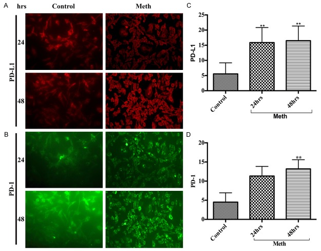
Figure 2.
A delayed increase of PD-L1 and PD-1 in hBECs. Representative immunocytochemical staining of (A) PD-L1 and (B) PD-1, in hBECs with/without METH at 24 hrs and 48 hrs; Ranks-Kruskal-Wallis test for quantitative densitometric analysis of (C) PD-L1 and (D) PD-1 levels. **P≤0.01 is the statistical significance compared with controls.

