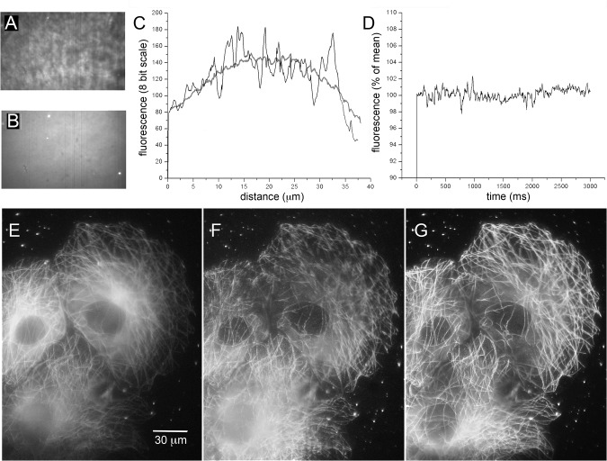
Fig 2. Improved field uniformity with shadowless TIRF excitation.
(A, B) Images (500x300 pixel, 40 x 24 μm) of rhodamine film on a coverglass. The image in A was captured using stationary-spot (conventional) TIRF excitation at 488 nm; that in B shows the same specimen but imaged by spinning-spot TIRF excitation. Dirt specks and fluorescent beads are readily discerned in B, but are almost completely masked by irregularities in the excitation field in A. The grey-scale levels in each panel were adjusted to equalize black levels and the brightest regions (excluding the fluorescent beads in B). (C) Measurements of fluorescence intensity along horizontal lines drawn across the center of the images in A (irregular black trace) and B (grey trace). (D) Stability of fluorescence excitation over time. The plot shows mean fluorescence intensity within a 100 x 100 pixel region in the center of the field, measured from 600 image frames acquired at 5 ms intervals in spinning-spot TIRF mode. (E-G) Representative images showing COS-7 cells expressing tubulin tagged by EGFP, captured using wide-field excitation (E), by stationary-spot TIRF (F) and spinning-spot TIRF (G). Images were captured with 488 nm excitation during 2s exposures using an 18 Mpixel Canon EOS M camera and eyepiece adapter.

