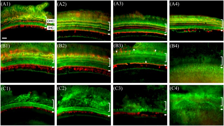
Fig 4. Whole-mounts of the auditory epithelium in normal (A1, A2, A3 and A4), cochleostomy only (B1, B2, B3 and B4) and blood injected ear (C1, C2, C3 and C4) at 2 weeks after surgery stained for myosin VIIa (red) for hair cells and rhodamine phalloidin (green) for actin and photographed with epifluorescence.
Hair cells loss was more severe in blood injected ear (C1~C4) than control ear (B1~B4). In blood injected ear, severe outer hair cell loss is observed from the basal turn up to the apex (C1~C4). A1, B1 and C1: Apical turn, A2, B2 and C2: 3rd turn, A3, B3 and C3: 2nd turn, A4, B4 and C4: basal turn, OHC: outer hair cell, IHC: inner hair cell. Scale bar = 30 μm.

