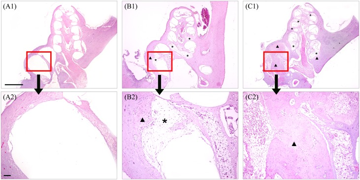
Fig 6. Sectional histopathologic findings of cochlea in normal (A), cochleostomy only ear (B) and blood injected ear (C) at 2 months after surgery.
More extensive fibrosis (asterisk) and ossification (arrow head) were observed in blood injected ear (C1 and C2) than control ear (B1 and B2). Scale bar = 500 μm in A1, B1 and C1 and 50 μm in A2, B2 and C2.

