
Fig. 3.
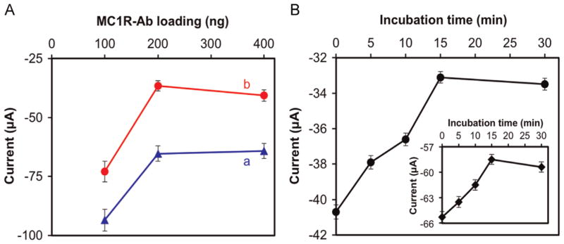
Peak anodic current responses of the immunosensor as a function of (A) amount of MC1R-Abs loaded and incubated for 15 min with (a) 2500 SK-MEL-2 cells/2.5 mL, and (b) 7500 SK-MEL-2 cells/2.5 mL. (B) The immunosensor was incubated in 7500 SK-MEL-2 cells/2.5 mL under various incubation times. Inset in “B” is the response for 2500 SK-MEL-2 cells/2.5 mL. Experimental conditions: 1 × PBS solution (pH 7.4) containing 1 mM [Fe(CN)6] 3−, 0.1 M KCl, and 100 μM EDTA at room temperature; scan rate=50 mV s−1. Error bars represent standard deviations of three independent measurements. detect and enumerate melanoma cells.