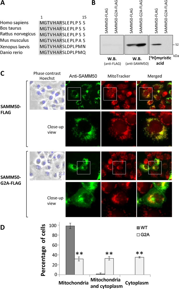
Fig 4. Protein N-myristoylation is required for proper targeting of SAMM50 to mitochondria.
A. Interspecies alignment of the N-terminal sequences of SAMM50. N-myristoylation motifs are shown in grey in the N-terminal sequence. B. Detection of protein N-myristoylation of SAMM50 expressed in COS-1 cells. cDNAs encoding SAMM50-FLAG and SAMM50-G2A-FLAG were transfected into COS-1 cells, and their expression and the N-myristoylation of the products in total cell lysates were evaluated by western blotting analysis (using anti-FLAG or anti-SAMM50 antibodies) and [3H]myristic acid labeling, respectively. C. Intracellular localization of SAMM50-FLAG and SAMM50-G2A-FLAG was determined by immunofluorescence staining of COS-1 cells transfected with cDNAs encoding these two proteins using an anti-SAMM50 antibody. Mitochondria were detected using MitoTracker Red. Lower panels show a close-up image of the area outlined by a white box in the upper panels. D. Quantitative analysis of the intracellular localization of SAMM50-FLAG and SAMM50-G2A-FLAG. cDNAs encoding SAMM50-FLAG and SAMM50-G2A-FLAG were transfected into COS-1 cells and the intracellular localization of the expressed proteins in each cell was determined by immunofluorescence staining, and the extent of mitochondrial localization was compared. Quantitative analysis of the mitochondrial localization of SAMM50-FLAG and SAMM50-G2A-FLAG was performed by fluorescence microscopic observation of 50 immunofluorescence-positive (transfected) cells. The extent of mitochondrial localization was expressed as a percentage of the number of cells in which selective localization to mitochondria, localization to mitochondria and cytoplasm, and selective localization to cytoplasm was observed against the total number of transfected cells. Data are expressed as mean ± SD for five independent experiments. **P < 0.001 vs. wild-type.

