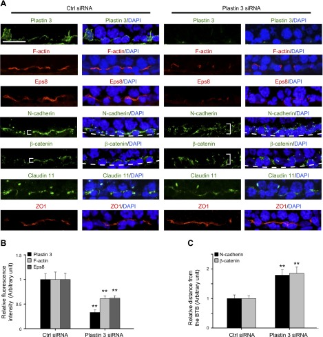
Figure 7.
Changes in the distribution of F-actin, actin regulatory protein Eps8, and basal ES proteins following a KD of plastin 3 in adult rat testes in vivo. Frozen sections of rat testes transfected with siRNA duplexes to knock down plastin 3 vs. nontargeting negative control duplexes were stained for plastin 3, F-actin, actin barbed end capping and bundling protein Eps8, as well as basal ES proteins N-cadherin and β-catenin vs. TJ proteins claudin-11 and ZO-1. Cell nuclei were visualized with DAPI (blue). Stage VII/VIII/IX tubules were randomly selected for analysis. A) KD of plastin 3 was verified in these tubules in which plastin 3 fluorescence staining was considerably diminished. The fluorescence intensity of both F-actin and Eps8 at the BTB was also weakened after plastin 3 KD vs. control, supporting the notion that there were less F-actin and microfilament bundles in plastin 3 KD testes. Consistent with the findings in vitro, the basal ES proteins N-cadherin and β-catenin were no longer restrictively localized to the BTB near the basement membrane (annotated by dashed white line); instead, they displayed a considerably diffused pattern (see white brackets in plastin 3 KD testes vs. control testes). Interestingly, the TJ proteins claudin-11 and ZO-1 were not affected by the plastin 3 KD. Scale bar, 40 µm. B and C) Semiquantitative analysis of fluorescence image data shown in (A) including fluorescence intensity (B) or the mislocalization of basal ES proteins by diffusing away from the BTB (C). Data in the control group were arbitrarily set at 1. Each bar is a mean ± sd of 3 rats. **P < 0.01.

